Slime Molds (Myxomycetes), Part 3
Lycogala to Trichia
Go to: Slime Molds, Part 4
Back to: Slime Molds Index
Slime Molds (Myxomycetes), Part 3 Go to: Slime Molds, Part 4 |
|
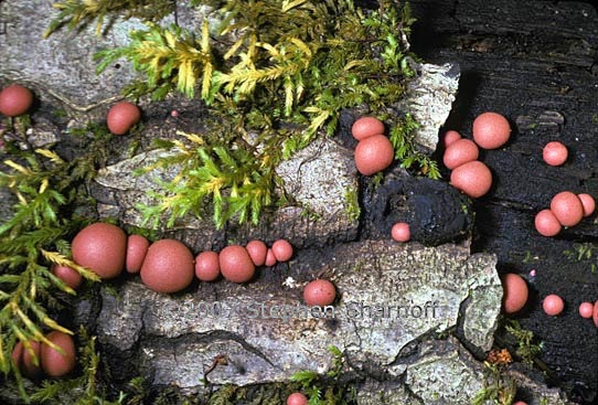
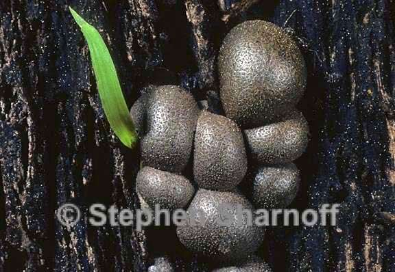
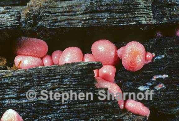
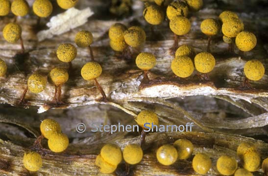
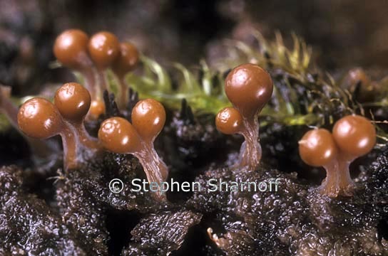
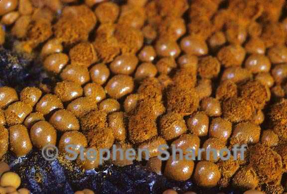
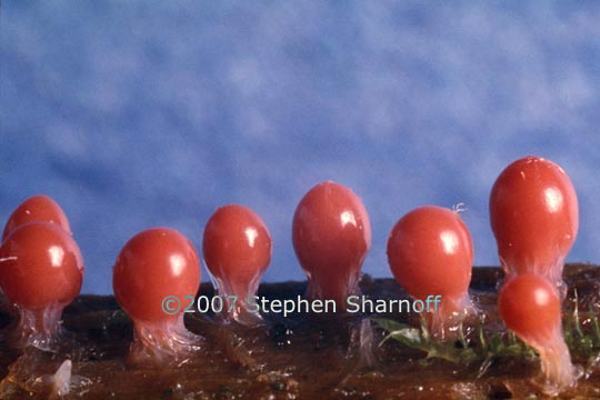
Image 1. Sporangia of Lycogala epidendrum
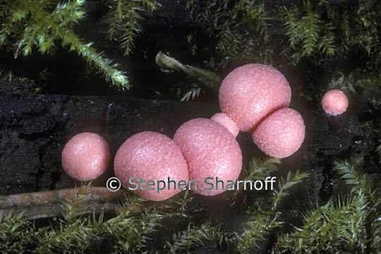
Image 2. Sporangia of Lycogala epidendrum
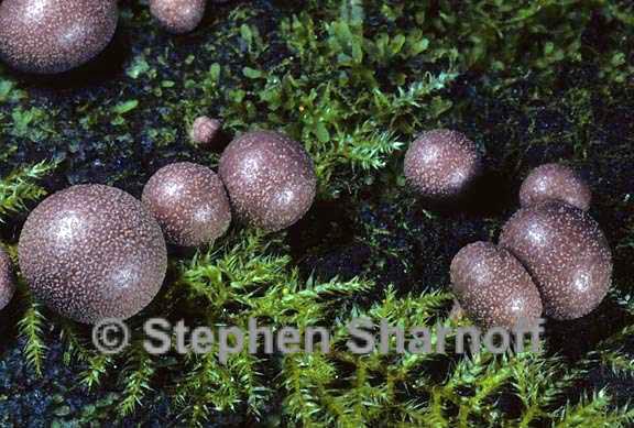
Image 3. Sporangia of Lycogala epidendrum
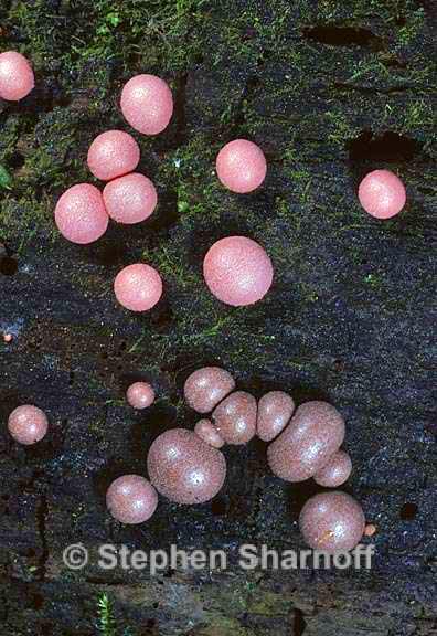
Image 4. Sporangia of Lycogala epidendrum
Image 5. Sporangia of Lycogala epidendrum
Image 6. Sporangia of Lycogala epidendrum
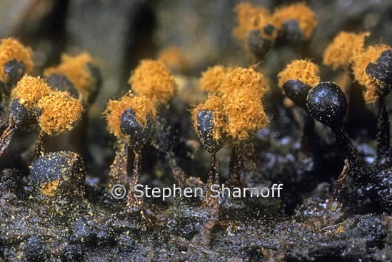
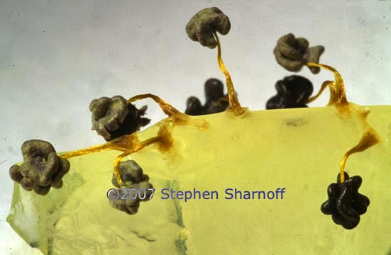
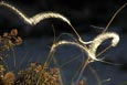
Image 7. Metatricha floriformis
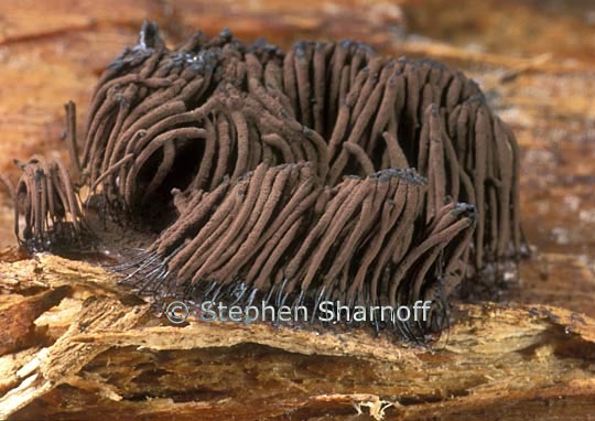
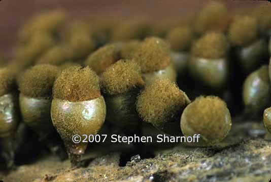
Image 8. Metatrichia vesparium, from southern Ontario
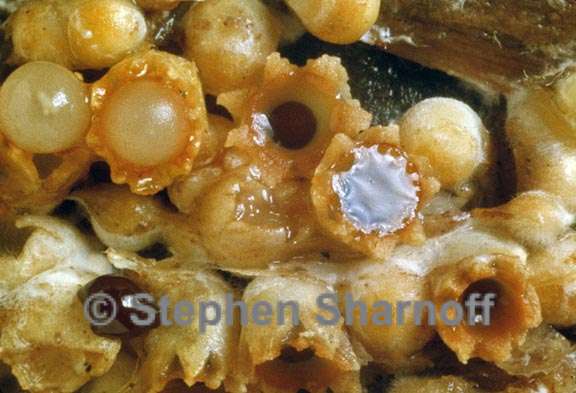
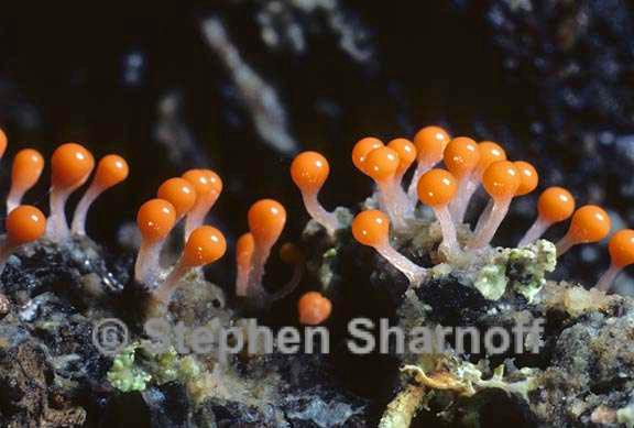
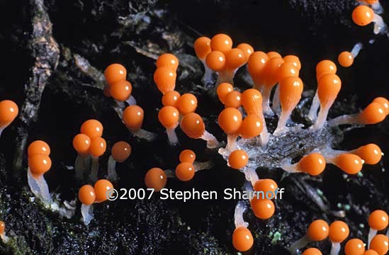
Image 9. Possibly Physarum flavicomum, from near Prince George, British Columbia
Image 10. Physarum polycephalum sporangia on agar, a laboratory growth medium
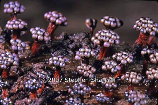
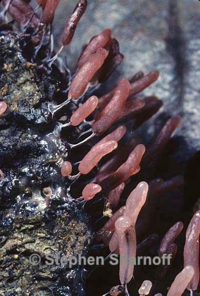
Image 11. Prototrichia metallica, from the subalpine Sierra Nevada, California
Image 12. Sphaerobolus sp.from Tilden Park, near Berkeley, California
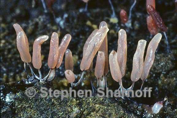
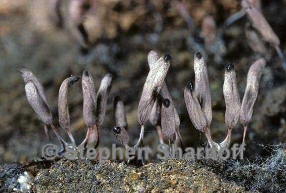
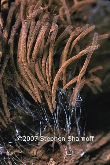
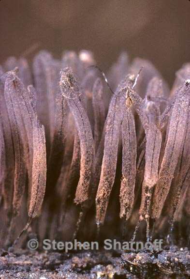
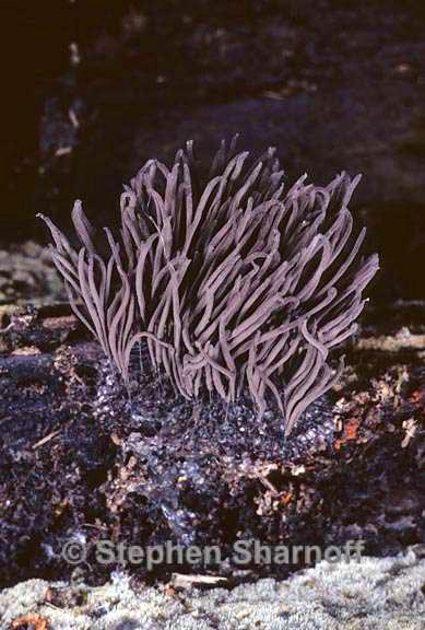
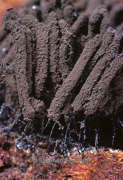
Image 13. Steminotopsys typhina
Image 14. Steminotopsys typhina
Image 15. Steminotopsys typhina
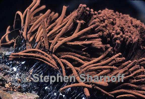
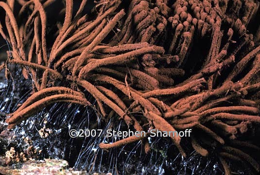
Image 16. Stemonitis axifera
Image 17. Stemonitis axifera
Image 18. Stemonitis axifera
Image 19. Stemonitis axifera
Image 20. Stemonitis sp. Most of the spores have been released.
Image 21. Stemonitis sp. Location uncertain
Image 22. Stemonitis sp. Location uncertain
Image 23. Trichia decipiens, from near Stewarts Point, Sonoma County, California
Image 24. Trichia decipiens, from near Stewarts Point, Sonoma County, California
Image 25. Possibly Trichia decipiens
Image 26. Immature sporangia, possibly a species of Trichia
Image 27. Trichia sp., from Humboldt County, northwestern California.
Image 28. Trichia sp. |
| Go to: Slime Molds, Part 4 | Back to: Slime Molds Index |